Superior tarsal muscle
| Superior tarsal muscle | |
|---|---|
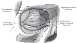
 The tarsi and their ligaments. Right eye; front view (muscle not labeled but region is visible). | |
 Sagittal section of right orbital cavity (muscle not labeled but region is visible). | |
| Details | |
| Origin | Underside of levator palpebrae superioris |
| Insertion | Superior tarsal plate of the eyelid |
| Artery | Ophthalmic artery |
| Nerve | Sympathetic nervous system |
| Actions | Raises the upper eyelid |
| Identifiers | |
| Latin | musculus tarsalis superior |
| TA98 | A15.2.07.045 |
| TA2 | 6828 |
| FMA | 49058 |
| Anatomical terms of muscle | |
The superior tarsal muscle is a smooth muscle adjoining the levator palpebrae superioris muscle muscle that helps to raise the upper eyelid.
Structure
[edit]The superior tarsal muscle originates on the underside of levator palpebrae superioris muscle and inserts on the superior tarsal plate of the eyelid.
Nerve supply
[edit]The superior tarsal muscle receives its innervation from the sympathetic nervous system. Postganglionic sympathetic fibers originate in the superior cervical ganglion, and travel via the internal carotid plexus, where small branches communicate with the oculomotor nerve as it passes through the cavernous sinus.[1] The sympathetic fibres continue to the superior division of the oculomotor nerve, where they enter the superior tarsal muscle on its inferior aspect.
Function
[edit]Its role is not fully clear, but may be an accessory muscle to raise the upper eyelid.[2]
Clinical significance
[edit]Damage to some elements of the sympathetic nervous system can inhibit this muscle, causing a drooping eyelid (partial ptosis). This is seen in Horner's syndrome. The ptosis seen in Horner's syndrome is of a lesser degree than is seen with an oculomotor nerve palsy.
History
[edit]The muscle derives its name from Greek ταρσός 'flat surface', typically used for drying.
The term Müller's muscle is sometimes used as a synonym.[3] However, the same term is also used for the circular fibres of the ciliary muscle,[4][5] and also for the orbitalis muscle that covers the inferior orbital fissure. Given the possible confusion, the use of the term Müller's muscle should be discouraged unless the context removes any ambiguity.
See also
[edit]References
[edit]- ^ Snell R, Lemp M (1998). Clinical Anatomy of the Eye (2nd ed.). Oxford, UK: Blackwell Publishing. ISBN 9780632043446.[page needed]
- ^ Standring, S, ed. (2021). "Orbit and accessory visual apparatus". Gray's anatomy : the anatomical basis of clinical practice (42 ed.). New York. ISBN 978-0-7020-7705-0.
{{cite book}}: CS1 maint: location missing publisher (link) - ^ van der Werf F, Baljet B, Prins M, Timmerman A, Otto JA (June 1993). "Innervation of the superior tarsal (Müller's) muscle in the cynomolgus monkey: a retrograde tracing study". Investigative Ophthalmology & Visual Science. 34 (7): 2333–40. PMID 7685010.
- ^ doctor/2564 at Who Named It?
- ^ "Glossary of Eponyms". Retrieved 2008-02-23.
Further reading
[edit]- Beard C (April 1985). "Müller's superior tarsal muscle: anatomy, physiology, and clinical significance". Annals of Plastic Surgery. 14 (4): 324–33. doi:10.1097/00000637-198504000-00005. PMID 3994278. S2CID 44374772.
- Dortzbach RK (October 1979). "Superior tarsal muscle resection to correct blepharoptosis". Ophthalmology. 86 (10): 1883–91. doi:10.1016/S0161-6420(79)35341-6. PMID 399800.
- Felt DP, Frueh BR (1988). "A pharmacologic study of the sympathetic eyelid tarsal muscles". Ophthalmic Plastic and Reconstructive Surgery. 4 (1): 15–24. doi:10.1097/00002341-198801130-00003. PMID 2979003. S2CID 34606836.
- van der Werf F, Baljet B, Prins M, Ruskell GL, Otto JA (October 1996). "Innervation of the palpebral conjunctiva and the superior tarsal muscle in the cynomolgous monkey: a retrograde fluorescent tracing study". Journal of Anatomy. 189 ( Pt 2) (Pt 2): 285–92. PMC 1167745. PMID 8886950.
- Putterman AM, Urist MJ (August 1975). "Müller muscle-conjunctiva resection. Technique for treatment of blepharoptosis". Archives of Ophthalmology. 93 (8): 619–23. doi:10.1001/archopht.1975.01010020595007. PMID 1156223.
