Inflammaging

Inflammaging (also known as inflamm-aging or inflamm-ageing) is a chronic, sterile, low-grade inflammation that develops with advanced age, in the absence of overt infection, and may contribute to clinical manifestations of other age-related pathologies. Inflammaging is thought to be caused by a loss of control over systemic inflammation resulting in chronic overstimulation of the innate immune system. Inflammaging is a significant risk factor in mortality and morbidity in aged individuals.[2][3][4]
Inflammation is essential to protect against viral and bacterial infection, as well as noxious stimuli. It is an integral part of the healing process, though prolonged inflammation can be detrimental. The network dynamics of inflammation change with age, and factors such as genes, lifestyle, and environment contribute to these changes.[5] Current research studying inflammaging is focused on understanding the interaction of dynamic molecular pathways underlying both aging and inflammation and how they change with chronological age.
Characteristics
[edit]Fine control and modulation of the immune system response becomes fragile and less precise with age, as seen with other bodily systems.[5] Remodeling of the immune system in the elderly is thought to be characterized by an inability to control systemic inflammation. With age, the number of lymphocytes being produced decreases, and the composition and quality of the mature lymphocyte pool changes.[6] While the effectiveness of adaptive immune system declines, innate immune mechanisms become overactive and less precise, leading to an increase in pro-inflammatory phenotypes that contributes to "inflammaging."[5] The secretome of senescent mesenchymal stem and stromal cells (MSCs) exhibits immunomodulatory effects and serves as a driver in inflammaging-related diseases.[7][8] All together, this contribues to a less efficient immune system response to pathogens and chronic, systemic inflammatory phenotypes.
Causes
[edit]Inflammaging is a complex and systemic issue, likely a result of several factors.
Inflammasome activation
[edit]
Over-activation of the inflammasome is one mechanism contributing to inflammaging. The inflammasome is a multi-protein complex consisting of a sensor, an adapter, and an effector, that when activated, modulates caspases which cleave cytokines and result in an inflammatory signaling response.[9][10][11] Receptors present on the cell surface act as sensors for the detection of damage and pathogens. When activated, this system can go on to elicit pro-inflammatory cytokine secretion and sometimes cell death.[12][13] Pro-inflammatory cytokines secretion acts as the effector or the response to such stimuli.
Stimuli that can fuel inflammasome assembly include pathogen-associated molecular patterns (PAMPS), damage associated molecular patterns (DAMPS), nutrients, and the microbiota.[14] These various self, non-self, and quasi self molecules are recognized by receptors of innate immune cells, whose promiscuity allows for many different signals to lead to activation and consequently to inflammation. Examples of stimuli that act as PAMPS include viral and bacterial infection, such as cytomegalovirus and periodontitis, respectively. Examples of DAMPS include misfolded and oxidized proteins, cell debris, and self nucleic acids.
Generation of reactive oxygen species
[edit]In addition to inflammasome activation, with age, cellular components accumulate reactive oxygen species (ROS). These free radicals can damage DNA, lipid, and protein and is able to drive cellular senescence. This is accompanied by a loss in efficiency of DNA damage repair mechanisms.[15] This results in pro-inflammatory cytokine secretion, which contributes to low grade, chronic inflammation in the absence of pathogen or damage, but rather in response to damaged self molecules like oxidized nucleotides.[16]
SASP phenotype
[edit]Senescent cell populations increase with age and secrete a pro-inflammatory cocktail of chemicals, a condition known as senescence-associated secretory phenotype (SASP).[14] Cells with the SASP are characterized by being in cell cycle arrest, releasing inflammatory factors, and possessing a particular morphology. These cells promote tissue degeneration and are able to spread to other regions by way of the inflammatory secretory molecules released.[17] This contributes to inflammaging as inflammatory secretion contributes to innate immune activation and exhaustion.
Functional decline in autophagy and mitophagy
[edit]Another contribution to inflammaging is a decline in effective autophagy and mitophagy capacity. This is an essential process for cellular housekeeping that prevents protein aggregation and accumulation of damaged mitochondria that produce large quantities of reactive oxygen species.[18] A loss of effective autophagic processes leads to aggregation of damaged proteins. As inflammasome precision declines with age, these aggregates, normally degraded, can be recognizes as a pathogen and lead to an inflammatory response. This contributes to inflammaging and is also involved in many neurodegenerative diseases as well.
Other factors
[edit]Other possible factors that may lead to inflamm-aging include insufficient sleep, overnutrition, sensory overload, physical inactivity, altered gut microbiome, impaired intestinal epithelial barrier, and chronic stress occurring in any stage of the individual's life.[19][20][21] Cytokines with inflammatory properties can also be secreted by fat tissue.[22]
Biomarkers of inflammaging
[edit]
Cytokines are currently used as biomarkers of inflammaging as they are indicative of inflammation and play a large role in the regulation of pro and anti-inflammatory immune regulation. Cytokines are small proteins that are secreted by many cell types that are very relevant in the study of aging and longevity. Aging studies show that a healthy balance of pro and anti-inflammatory cytokine secretion is associated with successful aging whereas dysregulation of this system results in inflammaging, poor aging phenotypes, and other aging-related diseases.[2] Currently, levels of TNFa, IL-6 and IL-1 can be used as inflammatory biomarkers that indicate frailty, an altered immune system, functional decline and mortality associated with inflammaging .[23]
Biomarkers of immuno-senescence also exist and involved changes to T cells, CD4/CD8 ratios as well as the SASP phenotype. Altogether, these biomarkers may not be translationally relevant to clinical outcomes. The generation of more reliable biomarkers of inflammation and aging is of interest in current research.
Interleukin 6
[edit]IL-6 is pro-inflammatory in nature and can be produced by many cells of the immune system as well as non-immune cells, like fibroblasts.[24] This cytokine has been identified in many organs such as the lungs, adipose tissue, muscle, and brain. The concentration of this cytokine is usually very low or non-detectable in young adults though levels increase in old age and are very high in the elderly.[2] Moreover, elevated IL-6 has also been associated with disability and mortality in older adults. High serum levels are associated with cognitive impairment, low locomotion, and depression.[24]

Interleukin 1
[edit]The interleukin 1 family consists of both pro and anti inflammatory mediators and there are 9 genes that encode different forms of interleukin 1, like Il-1a and IL-1B.[25] IL-1B is one of the most prominent mediators of inflammation and its secretion is tightly regulated given its potent nature.[9] IL-1B starts in an inactive form and is induced first by toll like receptors or TNFa and requires a second stimulus by the inflammasome to induce the mature, active form of IL-1B.[9]
Tumor necrosis factor-alpha
[edit]Tumor necrosis factor-alpha (TNF-alpha) is an inflammatory cytokine produced upon acute inflammation and is an important signaling molecule responsible for inducing apoptosis or necrosis.[26] TNF-alpha exerts its effects by binding to various membrane receptors that belong to the TNF Receptor superfamily. With age, TNF-alpha serum levels negatively correlate with T cell function.[27] Additionally, elevated TNF-alpha levels are associated with increased systemic inflammation and contribute to inflammatory diseases like rheumatoid arthritis.[27] TNF-alpha signaling is thought to be up-regulated during inflammaging and contributes to cellular senescence and immune exhaustion.
Evolutionary consideration
[edit]While inflammation is capable of having negative implications, evolutionarily insight explains how inflammation has served as a layer of protection. It has been proposed that immune, metabolic, and endocrine systems co-evolved.[28]
In prehistoric times, starvation and infection by a pathogen pose as severe risks to survival. Inflammation may have served a protective role in human survival when food and water were scarce and highly contaminated. This explains post prandial inflammation, which involves innate immune system activation after ingesting a meal. Additionally, during infection by a pathogen, leptin synthesis changes and a reduction in food intake occurs.[28] This is to decrease the likelihood of ingesting another pathogen as well as preserving receptors critical for pathogen sensing, from sensing pathogenic nutrients instead. Perhaps, this is another reason calorie restriction is beneficial in the treatment of inflammaging.[28]

These same inflammatory processes may be detrimental towards humans in current society where over-nutrition is readily available. While inflammatory adaptations have evolved to promote survival in times of food deprivation, it does not appear that such adaptations have evolved in periods of over-nutrition.[29] In current times, natural selection does not favor those who are spared from inflammaging, as this occurs at ages past the reproduction window.
Inflammaging and COVID-19
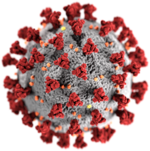
[edit]Adaptive immunity, involving the ability to fight off pathogens, declines with age.[30] Chronic inflammation and immunosenescence, which both increase with advanced chronological age, renders the elderly population more vulnerable to adverse, long term effects of viral infection by SARS-CoV2.[31] Inflammaging alone contributes to pro-inflammatory cytokine secretion that, in combination with viral infection by SARS-CoV2, may exhaust immune system function, contributing to worse outcomes when fighting COVID-19.[31]

Available evidence indicates that SARS-CoV2 enters the central nervous system through the lymphatic system and the virus was confirmed present in the capillaries and neuronal cells of the frontal lobe of COVID-19 patients.[31] This is corroborated with evidence demonstrating that SARS-CoV2 was present in cerebral spinal fluid of infected patients which displayed severe neurological symptoms. Viral infection is capable of inducing neuroinflammation through neuro-immune interactions. While aging is the most significant risk factor in the development of neurodegenerative diseases like Alzheimers, Parkinson's, and Amyotrophic lateral sclerosis, chronic, low-grade inflammation and immunosenescence may be aggravated by a viral infection, worsening the aging phenotype and contributing to the development of neurodegenerative disease. For example, neuroinflammation has been shown to contribute greatly to the severity and pathogenesis of Parkinson's disease. Infection by the H1N1 virus was shown to contribute to Parkinson's disease development.[31]
Inflammaging and COVID-19 infection may lead to worse outcomes and contribute to the development of neurodegenerative disease in aged individuals.
Cardiovascular diseases
[edit]Inflammaging has been linked to a higher risk of cardiovascular diseases and has been increasingly recognized as a determinant of cardiovascular outcome.[32]
References
[edit]- ^ "BioRender". BioRender. Retrieved 29 November 2021.
- ^ a b c Alberro, Ainhoa; Iribarren-Lopez, Andrea; Sáenz-Cuesta, Matías; Matheu, Ander; Vergara, Itziar; Otaegui, David (23 February 2021). "Inflammaging markers characteristic of advanced age show similar levels with frailty and dependency". Scientific Reports. 11 (1): 4358. Bibcode:2021NatSR..11.4358A. doi:10.1038/s41598-021-83991-7. ISSN 2045-2322. PMC 7902838. PMID 33623057. S2CID 232039252.
- ^ Fulop, T., Larbi, A., Pawelec, G., Khalil, A., Cohen, A. A., Hirokawa, K., ... & Franceschi, C. (2021). Immunology of aging: the birth of inflammaging. Clinical reviews in allergy & immunology, 1-14. PMID 34536213 PMC 8449217 doi:10.1007/s12016-021-08899-6
- ^ Dugan, B., Conway, J., & Duggal, N. A. (2023). Inflammaging as a target for healthy ageing. Age and Ageing, 52(2), afac328. PMID 36735849 doi:10.1093/ageing/afac328
- ^ a b c Rea, Irene Maeve; Gibson, David S.; McGilligan, Victoria; McNerlan, Susan E.; Alexander, H. Denis; Ross, Owen A. (9 April 2018). "Age and Age-Related Diseases: Role of Inflammation Triggers and Cytokines". Frontiers in Immunology. 9: 586. doi:10.3389/fimmu.2018.00586. ISSN 1664-3224. PMC 5900450. PMID 29686666.
- ^ Montecino-Rodriguez E, Berent-Maoz B, Dorshkind K (March 2013). "Causes, consequences, and reversal of immune system aging". The Journal of Clinical Investigation. 123 (3): 958–65. doi:10.1172/JCI64096. PMC 3582124. PMID 23454758.
- ^ Lee, B.C.; Yu, K.R. (2020). "Impact of mesenchymal stem cell senescence on inflammaging". BMB Reports. 53 (2): 65–73. doi:10.5483/BMBRep.2020.53.2.291. ISSN 1976-670X. PMC 7061209. PMID 31964472.
- ^ Lyamina, S.; Baranovskii, D.; Kozhevnikova, E.; Ivanova, T.; Kalish, S.; Sadekov, T.; Klabukov, I.; Maev, I.; Govorun, V. (2023). "Mesenchymal Stromal Cells as a Driver of Inflammaging". International Journal of Molecular Sciences. 24 (7): 6372. doi:10.3390/ijms24076372. ISSN 1422-0067. PMC 10094085. PMID 37047346.
- ^ a b c Yazdi, Amir S.; Ghoreschi, Kamran (2016). "The Interleukin-1 Family". Regulation of Cytokine Gene Expression in Immunity and Diseases. Advances in Experimental Medicine and Biology. Vol. 941. pp. 21–29. doi:10.1007/978-94-024-0921-5_2. ISBN 978-94-024-0919-2. ISSN 0065-2598. PMID 27734407.
- ^ Latz, Eicke; Duewell, Peter (1 December 2018). "NLRP3 inflammasome activation in inflammaging". Seminars in Immunology. 40: 61–73. doi:10.1016/j.smim.2018.09.001. ISSN 1044-5323. PMID 30268598. S2CID 52890879.
- ^ Sebastian-Valverde, Maria; Pasinetti, Giulio M. (June 2020). "The NLRP3 Inflammasome as a Critical Actor in the Inflammaging Process". Cells. 9 (6): 1552. doi:10.3390/cells9061552. PMC 7348816. PMID 32604771.
- ^ Swanson, Karen V.; Deng, Meng; Ting, Jenny P.-Y. (August 2019). "The NLRP3 inflammasome: molecular activation and regulation to therapeutics". Nature Reviews Immunology. 19 (8): 477–489. doi:10.1038/s41577-019-0165-0. ISSN 1474-1733. PMC 7807242. PMID 31036962.
- ^ Malik, Ankit; Kanneganti, Thirumala-Devi (1 December 2017). "Inflammasome activation and assembly at a glance". Journal of Cell Science. 130 (23): 3955–3963. doi:10.1242/jcs.207365. ISSN 1477-9137. PMC 5769591. PMID 29196474.
- ^ a b Franceschi C, Campisi J (2014). "Chronic inflammation (inflammaging) and its potential contribution to age-associated diseases". Journal of Gerontology: Biological Sciences. 69 (Supp 1): s4–s9. doi:10.1093/gerona/glu057. PMID 24833586.
- ^ Chatterjee, Nimrat; Walker, Graham C. (June 2017). "Mechanisms of DNA damage, repair and mutagenesis". Environmental and Molecular Mutagenesis. 58 (5): 235–263. Bibcode:2017EnvMM..58..235C. doi:10.1002/em.22087. ISSN 0893-6692. PMC 5474181. PMID 28485537.
- ^ Zuo; Prather; Stetskiv; Garrison; Meade; Peace; Zhou (10 September 2019). "Inflammaging and Oxidative Stress in Human Diseases: From Molecular Mechanisms to Novel Treatments". International Journal of Molecular Sciences. 20 (18): 4472. doi:10.3390/ijms20184472. ISSN 1422-0067. PMC 6769561. PMID 31510091.
- ^ Gordleeva, Susanna; Kanakov, Oleg; Ivanchenko, Mikhail; Zaikin, Alexey; Franceschi, Claudio (1 October 2020). "Brain aging and garbage cleaning". Seminars in Immunopathology. 42 (5): 647–665. doi:10.1007/s00281-020-00816-x. ISSN 1863-2300. PMID 33034735. S2CID 222235623.
- ^ Salminen, Antero; Kaarniranta, Kai; Kauppinen, Anu (March 2012). "Inflammaging: disturbed interplay between autophagy and inflammasomes". Aging. 4 (3): 166–175. doi:10.18632/aging.100444. ISSN 1945-4589. PMC 3348477. PMID 22411934.
- ^ Franceschi C, Garagnani P, Parini P, Giuliani C, Santoro A (October 2018). "Inflammaging: a new immune-metabolic viewpoint for age-related diseases". Nature Reviews. Endocrinology. 14 (10): 576–590. doi:10.1038/s41574-018-0059-4. hdl:11585/675065. PMID 30046148. S2CID 50785788.
- ^ Meier J, Sturm A (2009). "The intestinal epithelial barrier: does it become impaired with age?". Digestive Diseases. 27 (3): 240–5. doi:10.1159/000228556. PMID 19786747. S2CID 40407420.
- ^ Kiecolt-Glaser JK, Preacher KJ, MacCallum RC, Atkinson C, Malarkey WB, Glaser R (July 2003). "Chronic stress and age-related increases in the proinflammatory cytokine IL-6". Proceedings of the National Academy of Sciences of the United States of America. 100 (15): 9090–5. Bibcode:2003PNAS..100.9090K. doi:10.1073/pnas.1531903100. PMC 166443. PMID 12840146.
- ^ Pedersen M, Bruunsgaard H, Weis N, Hendel HW, Andreassen BU, Eldrup E, Dela F, Pedersen BK (April 2003). "Circulating levels of TNF-alpha and IL-6-relation to truncal fat mass and muscle mass in healthy elderly individuals and in patients with type-2 diabetes". Mechanisms of Ageing and Development. 124 (4): 495–502. doi:10.1016/s0047-6374(03)00027-7. PMID 12714258. S2CID 20234017.
- ^ Fulop, Tamas; Cohen, Alan; Wong, Glenn; Witkowski, Jacek M.; Larbi, Anis (2019), Moskalev, Alexey (ed.), "Are There Reliable Biomarkers for Immunosenescence and Inflammaging?", Biomarkers of Human Aging, Healthy Ageing and Longevity, vol. 10, Cham: Springer International Publishing, pp. 231–251, doi:10.1007/978-3-030-24970-0_15, ISBN 978-3-030-24970-0, S2CID 203442315, retrieved 27 November 2021
- ^ a b Brábek, Jan; Jakubek, Milan; Vellieux, Fréderic; Novotný, Jiří; Kolář, Michal; Lacina, Lukáš; Szabo, Pavol; Strnadová, Karolína; Rösel, Daniel; Dvořánková, Barbora; Smetana, Karel (26 October 2020). "Interleukin-6: Molecule in the Intersection of Cancer, Ageing and COVID-19". International Journal of Molecular Sciences. 21 (21): 7937. doi:10.3390/ijms21217937. ISSN 1422-0067. PMC 7662856. PMID 33114676.
- ^ Kornman, Kenneth S. (February 2006). "Interleukin 1 genetics, inflammatory mechanisms, and nutrigenetic opportunities to modulate diseases of aging". The American Journal of Clinical Nutrition. 83 (2): 475S–483S. doi:10.1093/ajcn/83.2.475S. ISSN 0002-9165. PMID 16470016.
- ^ Idriss, H. T.; Naismith, J. H. (1 August 2000). "TNF alpha and the TNF receptor superfamily: structure-function relationship(s)". Microscopy Research and Technique. 50 (3): 184–195. doi:10.1002/1097-0029(20000801)50:3<184::AID-JEMT2>3.0.CO;2-H. ISSN 1059-910X. PMID 10891884. S2CID 3003943.
- ^ a b Frasca, Daniela; Blomberg, Bonnie B. (February 2016). "Inflammaging decreases adaptive and innate immune responses in mice and humans". Biogerontology. 17 (1): 7–19. doi:10.1007/s10522-015-9578-8. ISSN 1389-5729. PMC 4626429. PMID 25921609.
- ^ a b c Franceschi, Claudio; Garagnani, Paolo; Parini, Paolo; Giuliani, Cristina; Santoro, Aurelia (October 2018). "Inflammaging: a new immune–metabolic viewpoint for age-related diseases". Nature Reviews Endocrinology. 14 (10): 576–590. doi:10.1038/s41574-018-0059-4. hdl:11585/736269. ISSN 1759-5037. PMID 30046148. S2CID 50785788.
- ^ Franceschi, C.; Bonafè, M.; Valensin, S.; Olivieri, F.; De Luca, M.; Ottaviani, E.; De Benedictis, G. (June 2000). "Inflamm-aging. An evolutionary perspective on immunosenescence". Annals of the New York Academy of Sciences. 908 (1): 244–254. Bibcode:2000NYASA.908..244F. doi:10.1111/j.1749-6632.2000.tb06651.x. ISSN 0077-8923. PMID 10911963. S2CID 1843716.
- ^ Bartleson, Juliet M.; Radenkovic, Dina; Covarrubias, Anthony J.; Furman, David; Winer, Daniel A.; Verdin, Eric (September 2021). "SARS-CoV-2, COVID-19 and the aging immune system". Nature Aging. 1 (9): 769–782. doi:10.1038/s43587-021-00114-7. ISSN 2662-8465. PMC 8570568. PMID 34746804.
- ^ a b c d Domingues, Renato; Lippi, Alice; Setz, Cristian; Outeiro, Tiago F.; Krisko, Anita (29 September 2020). "SARS-CoV-2, immunosenescence and inflammaging: partners in the COVID-19 crime". Aging (Albany NY). 12 (18): 18778–18789. doi:10.18632/aging.103989. ISSN 1945-4589. PMC 7585069. PMID 32991323.
- ^ Puspitasari, Yustina M.; Ministrini, Stefano; Schwarz, Lena; Karch, Caroline; Liberale, Luca; Camici, Giovanni G. (18 May 2022). "Modern Concepts in Cardiovascular Disease: Inflamm-Aging". Frontiers in Cell and Developmental Biology. 10: 882211. doi:10.3389/fcell.2022.882211. ISSN 2296-634X. PMC 9158480. PMID 35663390.
 This article incorporates text from this source, which is available under the CC BY 4.0 license.
