Jump to content

英文维基 | 中文维基 | 日文维基 | 草榴社区

Portal:Medicine/Selected picture archive of 2007

From Wikipedia, the free encyclopedia

December 24, 2007 - December 31.

[edit]
Male nervous system in 3D.

Photo credit: Original uploader was 3dscience.

December 17, 2007 - December 24.

[edit]
Male reproductive organs in sagittal view.

Photo credit: Original uploader was 3dscience.

December 10, 2007 - December 17.

[edit]
Parts of the skin in 3D.

Photo credit: Original uploader was 3dscience.

December 3, 2007 - December 10.

[edit]
Human distal femur shot with a 510-grain lead Minié ball fired from a .58 caliber Springfield Model 1862 rifle.

Photo credit: National Institutes of Health, Health & Human Services

November 26, 2007 - December 3.

[edit]
Surgical incision in right shoulder, taken 7 days after surgery. Surgical staple

Photo credit: Llywrch

November 19, 2007 - November 26.

[edit]
The earliest illustration of tuberous sclerosis, with clusters of facial angiofibromas, displayed in Rayer's colour atlas of skin diseases published in 1835: "Traité des maladies de la peau".

Photo credit: Bibliothèque Interuniversitaire de Médecine - http://www.bium.univ-paris5.fr/histmed/medica/page?00584&p=82

November 12, 2007 - November 19.

[edit]
Factor Correspondence Analysis Comparing Different Individuals from European Ancestry Groups. The analysis was performed using 749 single nucleotide polymorphisms (SNPs) informative for European substructure (selected from a genome-wide panel of more than 5700 SNPs).

Photo credit: Seldin MF, Shigeta R, Villoslada P, Selmi C, Tuomilehto J, et al. (2006) European Population Substructure: Clustering of Northern and Southern Populations. PLoS Genet 2(9): e143 Fig. 4(b)

November 5, 2007 - November 12.

[edit]
Diagram of the human heart.

Photo credit: Original uploader was Yaddah

October 29, 2007 - November 5.

[edit]
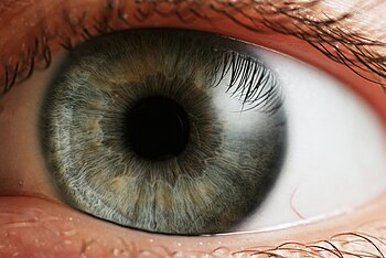
Photo credit: Original uploader was Che (CC-BY-2.5)

October 22, 2007 - October 29.

[edit]

Photo credit: Original uploader was 3dscience at en.wikipedia (CC-BY-2.5)

October 15, 2007 - October 22.

[edit]
Human heart.

Photo credit: User:Stanwhit607

October 8, 2007 - October 15.

[edit]
The image illustrates the processing principals of a positron emission tomograph (PET) commonly used during cancer diagnostics. It shows how during the annihilation process two photons are emitted in diametrically opposing directions. These photons are registered by the PET as soon as they arrive at the detector ring. After the registration, the data is forwarded to a processing unit which decides if two registered events are selected as a so-called coincidence event. All coincidences are forwarded to the image processing unit where the final image data is produced via image reconstruction procedures.

Photo credit: Public Domain

October 1, 2007 - October 8.

[edit]
The life cycle of Onchocerca volvulus, a parasitic worm which causes river blindness.

Photo credit: Giovanni Maki, derived from a CDC image at [1]

September 24, 2007 - October 1.

[edit]
The process of clinical islet transplantation for the treatment of diabetes mellitus.

Photo credit: Giovanni Maki through PLoS Medicine

September 17, 2007 - September 24.

[edit]
Behind the ear aid.

Photo credit: User:Nordelch

September 10, 2007 - September 17.

[edit]
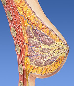
Breast normal anatomy cross-section view.

Photo credit: Patrick J. Lynch, medical illustrator

September 3, 2007 - September 10.

[edit]
Annotated diagram of cervical vertebrae from an oblique viewpoint.

Photo credit: user:debivort

August 27, 2007 - September 3.

[edit]
The Principle Organs and Vascular and Urino-Genital Systems of a Woman (c. 1507) is a drawing by Leonardo da Vinci.

Photo credit: Public domain

August 20, 2007 - August 27.

[edit]
SPECT nuclear imaging of the heart, short axis views.

Photo credit: Patrick J. Lynch, medical illustrator

August 13, 2007 - August 20.

[edit]
Heart posterior left ventricular wall infarction.

Photo credit: Patrick J. Lynch, medical illustrator

August 6, 2007 - August 13.

[edit]
Eye and orbit anatomy with motor nerves.

Photo credit: Patrick J. Lynch, medical illustrator

July 30, 2007 - August 6.

[edit]
Bronchial anatomy detail of alveoli and lung circulation.

Photo credit: Patrick J. Lynch, medical illustrator

July 23, 2007 - July 30.

[edit]
Artist's impression of the spatial representation of the human brain.

Photo credit: FSHL

July 16, 2007 - July 23.

[edit]
Cartoon of the alpha intercalated cell, showing the apical proton pump and the basolateral band 3 (kAE1)

Photo credit: public domain (by Felix-felix)

July 9, 2007 - July 16.

[edit]
Yersinia pestis seen at 2000x magnification. This bacterium, carried and spread by fleas, is generally thought to have been the cause of millions of deaths

Photo credit: public domain

July 2, 2007 - July 9.

[edit]
American Zouave ambulance crew demonstrating removal of wounded soldiers from the field, during the American Civil War.

Photo credit: Civil War glass negative collection (Library of Congress)

June 25, 2007 - July 2.

[edit]
CT scan showing lung cancer in the left lung.

Photo credit: Lange123

June 18, 2007 - June 25.

[edit]
Avian Flu vaccine development by reverse genetics techniques.

Photo credit: public domain

June 12, 2007 - June 18.

[edit]
Significant bilateral nephrocalcinosis (calcification of the kidneys) on a frontal X-ray (radiopacities (white) in the right upper and left upper quadrant of the image), as seen in distal renal tubular acidosis.

Photo credit: public domain

June 5, 2007 - June 12.

[edit]
Henipavirus structure

Photo credit: User:Rhys

May 28, 2007 - June 5.

[edit]
Pavlik harness is used to treat hip dysplasia.

Photo credit: User:Londenp

May 21, 2007 - May 28.

[edit]
Side-by-side comparison of two images of apples as seen by a trichromatic observer and the same apples as seen by a deuteranope (color blind individual)

Photo credit: User:Limbicsystem

May 14, 2007 - May 21.

[edit]
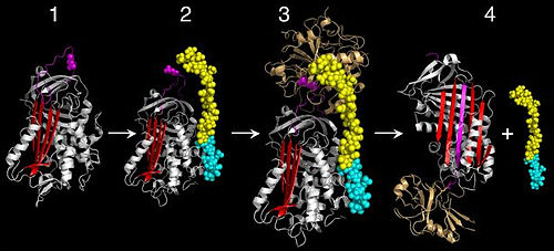
Serpin activity
Serpin activity
Serpin activity

Photo credit: Public domain

May 7, 2007 - May 14.

[edit]
A mature human spermatozoon

Photo credit: User:LadyofHats

April 30, 2007 - May 7.

[edit]

Photo credit: User:JHeuser

April 23, 2007 - April 30.

[edit]

Photo credit: User:Mjorter

April 16, 2007 - April 23.

[edit]
Herpes labialis. Cold sore on the lower lip. These infections may appear on the lips, nose or in surrounding areas. The sores may appear to be either weeping or dry, and may resemble a pimple, insect bite, or large chicken-pox lesion.

Photo credit: User:Metju

April 9, 2007 - April 16.

[edit]
Endoscopic image of deep gastric ulcer in the gastric antrum.

Photo credit: User:Samir

April 2, 2007 - April 9.

[edit]
A bone marrow harvest.

Photo credit: Public Domain

March 26, 2007 - April 2.

[edit]
Hand-drawing of the image of Subarachnoid Hemorrhage CT scan.

Photo credit: Chikumaya (ja.Wikipedia)

March 19, 2007 - March 26.

[edit]
File:Doctor and Child-by-David-Mason-flikr 5901759 9cfaa50cf7 o.jpg
"Doctor and Child" by David Mason

Photo credit: Flickr

March 12, 2007 - March 19.

[edit]
Picture of an Anthrax bacteria

Photo credit: Source

March 5, 2007 - March 12.

[edit]
Images from a cystoscopy. The top two images show the interior of a bladder. The top left image shows the bladder wall, the top right shows the cystoscope passing into the bladder from the urethra. The bottom two images show an inflamed urethra.

Photo credit: Cystoscopy carried out on Michael Reeve (29-year-old male), 25 April 2005, at the North London Nuffield Hospital

February 26, 2007 - March 12.

[edit]
Premature baby in an incubator. El Tahrir el Aam (General Liberation Hospital), Basrah, Iraq.

Photo credit: Thomas Hartwell

February 19, 2007 - February 26.

[edit]
A hysterosalpingogram. Note the catheter entering at the bottom of the screen, and the dark contrast material filling the uterine cavity (small triangle in the center) and outlining the Fallopian tubes (winding structures on left and right).

Photo credit: jemsweb

February 12, 2007 - February 19.

[edit]
Macrophages begin to fuse with, and inject its toxins into, the cancer cell. The cell starts rounding up and loses its spikes.

Photo credit: Susan Arnold (photographer)

February 5, 2007 - February 12.

[edit]
Insertion of an electrode during deep brain stimulation for Parkinson's disease.

Photo credit: User:Thomasbg

January 29, 2007 - February 5.

[edit]
Spiral CT depiction. Rotation of image acquisition apparatus (Tube and sensor array) around the patient.

Photo credit: Nevit Dilmen

January 22, 2007 - January 29.

[edit]
Acromion-clavicle disjonction (left shoulder) — note that the shoulder is lower and the "piano key"; the scar on the photograph and the screws on the radiography are ostheosynthesis material from a former trauma repair, without any connection with the present trauma

Photo credit: Christophe Dang Ngoc Chan

January 15, 2007 - January 22.

[edit]

Photo credit: Rakesh Ahuja, MD

January 8, 2007 - January 15.

[edit]
Photo of strabismus surgery. The medial rectus muscle is being disinserted following pre-placement of vicryl sutures. A Castroviejo locking forceps is grasping the superior pole of the muscle, while a pair of Manson-Aebli scissors does the cutting. The eyelids are being held by a Cook speculum.

Photo credit: User:Bticho

January 1, 2007 - January 7.

[edit]
Example of a partially completed computer-generated 3D facial reconstruction.

Photo credit: [2]