Talk:Two-photon excitation microscopy
| This article is rated C-class on Wikipedia's content assessment scale. It is of interest to the following WikiProjects: | ||||||||||||||||||||||||
| ||||||||||||||||||||||||
| The contents of the Multiphoton fluorescence microscope page were merged into Two-photon excitation microscopy on 22 April 2016. For the contribution history and old versions of the redirected page, please see its history; for the discussion at that location, see its talk page. |
,
A few inaccuarcies in the article
[edit]I would like to highlight a few aspects in the article that are wrong, inaccurate or difficult to understand and should be changed.
- "with 0.64 μm lateral and 3.35 μm axial spatial resolution" is correct for this specific study but is wrong in general. Higher resolution can be easily achieved with two-photon microscopy and depends on the wavelength and focusing NA of the excitation light.
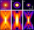
- "the multiphoton point spread function is typically dumbbell-shaped (longer in the x-y plane), compared to the upright rugby-ball shaped point spread function of confocal microscopes": This is simply wrong or at least misleading. The two microscope types generate a similar ellipsoid-shaped PSF.
- "Mode-locked Yb-doped fiber lasers with 325 fs pulses": reference is missing; also, I think it is not so relevant to be included in Wikipedia.
- Several references are a bit outdated (for current applications of two-photon imaging) and could be replaced.
- Some other sections, in particular in the beginning, are not very well written and could be improved in terms of structure and clarity.
If the authors of the article (or whoever feels responsible) agree, I would implement a few changes to improve the article. It would maybe be a good idea to take the german wikipedia article on this topic as a reference, because it is more organized and contains the relevant information. To be transparent, I'm working as a researcher (P.Rupprecht) in a lab employing and developing two-photon techniques (https://www.hifo.uzh.ch/en/research/helmchen/helmchenPeople.html). --89.206.81.76 (talk) 12:42, 31 March 2023 (UTC)
- Hi Peter,
- I agree with your points. More specifically, lateral and spatial resolution is probably not the information you need to have in the very first sentence on 2PEM.
- The examples of application are extremely short, rely on 1 - 2 references and are simply not informative regarding the value 2PEM provides in the fields. It might make sense to simply provide a list of fields in which the technique has been applied and give 1 - 2 more detailed examples.
- I checked the german version as you suggested and I agree that the structure is better (although the section on higher harmonic generation is taking just as much space as the part on 2P microscopy, which might be a bit too much - just personal taste, though).
- Maybe wait one or two days for additional feedback, but I, for one would welcome your changes.
- Best,
- Lukas Anneluka (talk) 13:14, 31 March 2023 (UTC)
- I've implemented the most important changes now. I deleted a few sentences which were out of context. It's still not a very good article, but I hope to have improved it a bit! Feedback and further suggestions are welcome! Ptrrupprecht (talk) 13:51, 3 April 2023 (UTC)
Extent of resolution improvement
[edit]I think the article would benefit if it's easy to add an example of the improvement of resolution, particularly z-direction over single-photon excitation, and comparison with confocal and other techniques.--Skoch3 03:28, 10 February 2007 (UTC)
i don't believe that z-resolution with multiphoton microscopy is improved. it should be somewhat worse, given that the the z-resolution is inversely related to the excitation wavelength. i believe it's roughly on the order of being 2x worse in the z-axis. i forget the details, but i believe you're correct in that this may be an important point to note.--cpschultz 01:20, 28 March 2007 (UTC)
the maximum z-resolution of a 2-photon is on the order of ~0.7 um, if memory serves... the thing that makes 2-photon resolution better is that there is virtually no excitation outside a very small volume, in 1-photon imaging, a much larger volume gets exited in the z because of scattering, etc... IlyaV (talk) 04:38, 30 December 2009 (UTC)
All of you are correct. The z-resolution in microscopy is depending on the focal volume of the illumination and the possibility to select the plane of interest in the "observation beam". While in confocal laser scanning microscopy the resolution is given almost exclusively by the latter (i.e. by the pinhole size), in 2p microscopy the focal volume determines the resolution. As a first estimate you can calculate the Rayleigh range of the beam of interest (emission in confocal, excitation in 2p), which is given by
In confocal microscopy, the emission wavelength is between 500 and 600nm (roughly), while in 2p, the excitation happens with 800 to 1000nm. So even if you assume that not the whole focal volume is excited in 2p microscopy, you still end up with about the same z-resolution (that is about 320 nm for a standard 1.0NA water immersion objective; twice the Rayleigh range). If you assume the whole focal volume is excited (over the complete Rayleigh range), z-resolution (and actually the lateral resolution as well) is worse in 2p than in confocal microscopy. Donik (talk) 20:03, 26 September 2011 (UTC)
From the article,
- The purpose of employing the two-photon effect is ... the resolution along the z dimension is improved, allowing for thin optical sections to be cut. ... the probability for fluorescent ... quadratically with the excitation intensity. ... Effectively, excitation is restricted to the tiny focal volume (~1 femtoliter), resulting in a high degree of rejection of out-of-focus objects.
Is this a mistake, i.e., isn't the idea to allow the sample to be cut into thicker sections without the usual associated sacrifice of resolving contrast? Cesiumfrog (talk) 23:31, 30 September 2014 (UTC)
energy conservation
[edit]how can two photons of 1/2E add up to be slightly more than E? should include explanation of this. —The preceding unsigned comment was added by 141.213.67.55 (talk) 21:16, 20 February 2007 (UTC).
when a fluorophore is excited, part of the energy is dissipated as heat (ie, a phonon is emitted,) as such, the energy of the emitted photon is less than the energy of the two photons. IlyaV (talk) 04:38, 30 December 2009 (UTC)
Images
[edit]I presume it is a mistake when the included figure states PTM. This should probably be changed to PMT (PhotoMultiPlier)?
[2014.03.17_20:55] Yes, the figure should indicate emission collection via photomultiplier tubes (PMTs).
While working on de:Multiphotonenmikroskop, I uploaded the following image files to commons, all from the same article. Just in case somebody feels like to work them into the English version as well... --Dietzel65 (talk) 22:14, 28 December 2008 (UTC)
Two verses multi
[edit]This article states that: "The two-photon excitation microscope is a special variant of the multiphoton fluorescence microscope." I am under the impression that two photon and multi photon are interchangeable terms with identical meanings and that one is not actually a "variant" of the other at all. I believe that the latter was used to avoid a trademark infringement. 2.220.208.183 (talk) 17:13, 30 July 2014 (UTC) — Preceding unsigned comment added by CMSINNOV (talk • contribs) 23:57, 15 September 2009 (UTC)
References
[edit]Reference 9 and 12 are duplicated. — Preceding unsigned comment added by 2.220.208.183 (talk) 17:12, 30 July 2014 (UTC)
 Fixed Thanks, benmoore 09:02, 31 July 2014 (UTC)
Present-day usage
[edit]It would be useful for visitors if the main article could explain whether the technique is still widely used or not. This probably requires someone who has expertise in this area to make some estimate, and perhaps add a few articles that reinforce that opinion. I, as a visitor and only semi-casual knowledgable person, right now wonder whether this technique from 1990 is still widely used today. I have not heard about it yet but I have seen a few laser scanning fluorescence microscopes so far (they often use multiple techniques, so it is hard to know what they all use and don't use, such as this technique or not). 2A02:8388:1641:8380:8920:7EEB:D50A:6466 (talk) 19:34, 9 October 2019 (UTC)
- C-Class Molecular Biology articles
- Unknown-importance Molecular Biology articles
- C-Class MCB articles
- Low-importance MCB articles
- WikiProject Molecular and Cellular Biology articles
- All WikiProject Molecular Biology pages
- C-Class physics articles
- Low-importance physics articles
- C-Class physics articles of Low-importance


